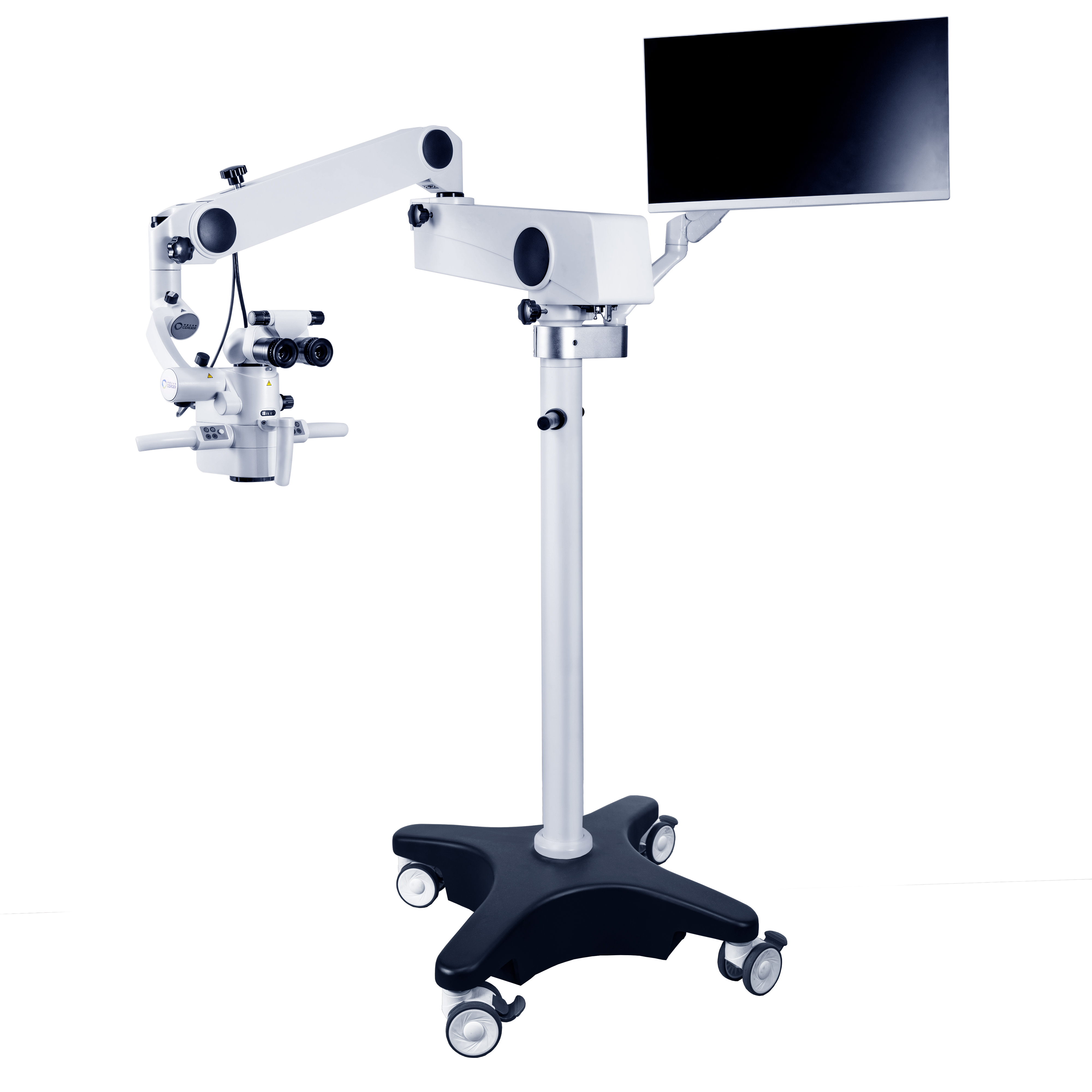
Optical magnification system: This is one of the core components of a microscope, like the lens of a camera, which determines the magnification and clarity of the image. The magnification of modern dental surgical microscopes is usually adjustable between 4-40 times, and doctors can easily switch the magnification according to the needs of the surgery, just like adjusting the camera focal length. Low magnification (4-8 times) is suitable for observing a large surgical field of view, such as viewing the overall condition of the surgical area during oral surgery; Medium magnification (8-14 times) meets the needs of most conventional dental surgeries, such as root canal treatment, periodontal surgery, etc.
Lighting system: Good lighting is the foundation for clear observation. The dental operating microscope adopts advanced lighting technology, such as LED cold light source, which can provide uniform, bright and shadow free light for the surgical area inside the oral cavity. This lighting method not only avoids the damage to oral tissue caused by the heat generated by traditional light sources, but also ensures that doctors can see every detail of the surgical site from any angle, just like performing on a bright stage, with every movement clearly visible.
Support and adjustment system: This system is like the “skeleton” and “joints” of a operating microscope, ensuring that the surgical microscope is stably placed in the appropriate position and can be flexibly adjusted. It can accurately adjust the height and angle according to the different needs of doctors and patients, allowing doctors to find the most comfortable and easy to observe position during operation, just like tailoring an exclusive operating platform for doctors.
Imaging and Recording System: Some high-end dental surgical microscopes are also equipped with imaging and recording systems, similar to a high-definition camera. It can display images under the Medical surgical microscope in real time on the screen, making it convenient for doctors to share observation results with assistants during the surgical process. At the same time, it can also record and take photos of the surgical process. These images and video materials can not only be used for subsequent case analysis and teaching research, but also allow patients to have a more intuitive understanding of their oral condition and treatment process.
The working principle of a dental microscope is based on the basic principles of optical imaging. Simply put, it magnifies small objects in the oral cavity through a combination of objective and eyepiece lenses. Light is emitted from the lighting system to illuminate the surgical area. The reflected light from the object is first magnified by the objective lens, then further magnified by the eyepiece, and finally forms a clear magnified image in the doctor’s eyes or on the imaging device. This is like using a magnifying glass to observe objects, but the magnification effect of a Oral surgery microscope is more precise and powerful, allowing doctors to see subtle details that are difficult for the naked eye to detect.

Media Contact
Company Name: Chengdu Corder Optics And Electronics Co., Ltd.
Email: Send Email
Country: China
Website: https://www.vipmicroscope.com/
